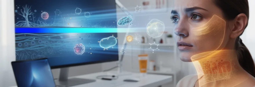
Modern life has fundamentally altered our relationship with light exposure. Where previous generations experienced primarily natural sunlight cycles, today’s digital landscape subjects our skin to unprecedented levels of artificial blue light radiation. This high-energy visible light, emitted by smartphones, computers, tablets, and LED lighting systems, penetrates deeper into dermal tissue than traditional UV radiation, triggering complex photobiological processes that accelerate skin aging. Recent dermatological research reveals that prolonged exposure to blue light generates reactive oxygen species, disrupts cellular repair mechanisms, and contributes to premature photoaging through mechanisms distinct from solar ultraviolet damage.
The proliferation of digital devices has created a new category of environmental skin stressors that dermatologists and skincare professionals must address. Blue light exposure now represents a significant factor in contemporary skin aging patterns, with studies indicating that average screen time exceeding 10 hours daily significantly impacts dermal health. Understanding these mechanisms becomes crucial for developing effective protection strategies that complement traditional photoprotection protocols.
Blue light spectrum analysis: understanding HEV wavelengths and photobiological mechanisms
The electromagnetic spectrum encompasses various wavelengths of light, each with distinct biological effects on human tissue. Blue light occupies the high-energy visible (HEV) portion of this spectrum, characterized by wavelengths ranging from 380 to 500 nanometers. This positioning places blue light immediately adjacent to ultraviolet radiation, sharing similar high-energy properties that enable deep tissue penetration and cellular interaction.
High-energy visible light properties: 380-500nm wavelength characteristics
Within the blue light spectrum, wavelengths between 380 and 400 nanometers demonstrate the highest photobiological activity, exhibiting properties that rival UV-A radiation in terms of dermal penetration depth. These shorter wavelengths carry significantly more energy per photon compared to longer visible light wavelengths, enabling them to interact directly with cellular components and trigger photochemical reactions within dermal tissue. The energy density of blue light at 415 nanometers, commonly emitted by LED screens, measures approximately 2.95 electron volts per photon, sufficient to induce molecular changes in skin proteins and lipids.
Research demonstrates that blue light penetration extends beyond the epidermis, reaching dermal layers where collagen and elastin fibers reside. This deep penetration capability distinguishes HEV light from other visible wavelengths, which typically affect only superficial skin layers. The photon energy at these wavelengths activates specific chromophores within skin cells, including flavins, porphyrins, and melanin precursors, initiating cascading photochemical reactions that ultimately manifest as visible skin aging.
Phototoxic reactions and reactive oxygen species generation in dermal tissue
Blue light exposure triggers immediate phototoxic responses within skin cells through the generation of reactive oxygen species (ROS). These highly unstable molecules, including singlet oxygen, hydroxyl radicals, and superoxide anions, interact aggressively with cellular components, causing oxidative damage to DNA, proteins, and lipid membranes. Oxidative stress resulting from blue light exposure exceeds the skin’s natural antioxidant capacity, leading to cumulative cellular damage that accelerates the aging process.
The mechanism of ROS generation involves the absorption of blue light photons by endogenous photosensitizers within skin cells. When these molecules absorb blue light energy, they transition to excited states, subsequently transferring this energy to molecular oxygen, creating reactive species. Studies indicate that one hour of blue light exposure from digital devices generates ROS levels comparable to 20 minutes of direct sunlight exposure, highlighting the significant oxidative burden imposed by modern technology use.
Circadian rhythm disruption effects on skin cellular repair mechanisms
Beyond direct photochemical damage, blue light exposure disrupts circadian rhythms that govern skin cell regeneration and repair processes. The skin operates on a 24-hour circadian cycle, with peak repair activity occurring during nighttime hours when melatonin production increases and cellular metabolism shifts toward restorative functions. Circadian disruption from evening blue light exposure suppresses melatonin synthesis, reducing the skin’s ability to repair daily damage and maintain
antioxidant defenses. Over time, this mismatch between day–night signaling and actual light exposure can translate into slower wound healing, diminished barrier recovery, and more pronounced fine lines. Experimental data show that keratinocytes exposed to blue light at night express altered clock genes, which in turn modify DNA repair and cell division timing. For you, that means late-night scrolling is not just stealing your sleep; it is also interfering with the precise schedule your skin relies on to correct daytime damage.
Melatonin, often called the “sleep hormone,” also functions as a powerful cutaneous antioxidant. Evening blue light suppresses melatonin not only systemically but also within the skin itself, reducing its capacity to neutralize free radicals generated during the day. This dual effect—less sleep and less local antioxidant activity—creates a compounding scenario where skin aging processes quietly accelerate. When we repeatedly expose our skin to blue light in the hours meant for repair, we are essentially asking our cells to work a night shift under fluorescent bulbs instead of in the dark, restorative environment they evolved for.
Digital device emission profiles: LED screens, OLED displays, and fluorescent lighting
Not all light-emitting technologies expose your skin to blue light in the same way. LED screens, which dominate smartphones, laptops, and modern monitors, typically have a pronounced emission peak around 440–460 nm, squarely within the high-energy visible light range implicated in oxidative stress. OLED displays, found in many newer premium devices, can be tuned to emit slightly less intense blue light at a given brightness, but they still contribute meaningfully to cumulative exposure, especially when viewed at close range.
Fluorescent lamps and certain high-efficiency LED lighting systems used in offices and homes also emit strong blue light components, often in the 400–470 nm range. While the intensity per unit area may be lower than direct sunlight, the long exposure times—eight to ten hours under overhead lighting—create a substantial total blue light dose. Studies comparing emission spectra show that a full workday in a brightly lit office environment plus evening screen use can rival, in HEV dose, several hours of midday outdoor exposure, even if the UV component is absent.
It is important to note that the skin experiences these artificial sources at much closer distances than the sun. A smartphone held 20–30 cm from the face focuses blue light on a relatively small skin area, particularly the periorbital region and cheeks. Over years, this localized, repetitive exposure pattern may contribute to specific aging signatures—such as periorbital hyperpigmentation or fine crow’s feet—that we increasingly see in high-screen-time populations. Understanding your personal “blue light environment” across devices and lighting is the first step toward realistic skin protection.
Dermatological pathophysiology of blue light-induced photoaging
Blue light-induced photoaging involves a network of molecular events that extend far beyond simple surface dryness or temporary redness. Once HEV photons penetrate to the epidermal and upper dermal layers, they interact with chromophores, generate reactive oxygen species, and trigger complex signaling pathways in keratinocytes and fibroblasts. These pathways converge on structural proteins such as collagen and elastin, pigment-producing melanocytes, and the microcirculation that supports skin vitality.
From a dermatological standpoint, blue light can be viewed as a chronic, low-grade stressor. It does not typically cause acute burns like UVB but instead drives subtle biochemical shifts that, over months and years, manifest as wrinkle formation, loss of elasticity, dullness, and stubborn hyperpigmentation. Several mechanistic pillars underlie this process: matrix metalloproteinase activation, melanogenesis dysregulation, mitochondrial impairment, and pro-inflammatory transcription factor activation. Together, they form the core of blue light-related skin aging.
Matrix metalloproteinase activation and collagen degradation pathways
One of the most critical consequences of blue light exposure is the upregulation of matrix metalloproteinases (MMPs), particularly MMP-1, which targets type I collagen. When ROS levels rise following HEV exposure, they activate signaling cascades that stimulate fibroblasts to produce these collagen-degrading enzymes. Over time, this enzymatic activity weakens the dermal extracellular matrix, leading to reduced firmness, increased laxity, and the fine lines we associate with digital-age photoaging.
Several in vitro studies have documented a significant increase in MMP expression after relatively short blue light exposure intervals, sometimes as brief as one hour. The effect is cumulative, meaning repeated low-dose exposures can be as detrimental as less frequent, higher-intensity insults. You can think of this as “micro-erosion” of your skin’s scaffolding: each session at your screen removes a tiny fraction of structural support, and the loss becomes visible only once enough of these micro-events have accumulated.
Compounding the issue, blue light simultaneously downregulates collagen synthesis pathways in fibroblasts by altering transforming growth factor-beta (TGF-β) signaling. This combination of increased breakdown and reduced production shifts the balance decisively toward net collagen loss. Clinically, this can present as earlier onset of nasolabial folds, finer but more numerous periorbital wrinkles, and a general loss of bounce in the mid-face, even in individuals who are otherwise diligent with UV-focused sun protection.
Melanogenesis dysregulation and hyperpigmentation formation mechanisms
Blue light also exerts a distinct influence on melanocytes, the pigment-producing cells within the basal epidermis. Unlike UV-induced melanogenesis, which is largely driven by DNA photodamage, HEV-associated pigmentation is closely linked to oxidative stress and photoreceptor-mediated signaling. Research has shown that blue light can activate opsin-3, a light-sensitive receptor expressed on melanocytes, leading to increased intracellular calcium and upregulation of melanogenic enzymes such as tyrosinase.
This signaling cascade results in a more sustained and often uneven melanin production response. In clinical settings, individuals with Fitzpatrick skin types III–VI appear particularly susceptible, developing longer-lasting hyperpigmented patches and exacerbations of melasma when exposed to repeated blue light. Unlike transient tanning, these HEV-related pigment changes may persist for months and can prove resistant to standard brightening treatments if ongoing exposure is not addressed.
Blue light-induced melanogenesis also tends to create a mottled, spotty appearance, especially on the cheeks, forehead, and periorbital area where screens are most frequently directed. If you already struggle with post-inflammatory hyperpigmentation from acne or previous sun damage, cumulative blue light exposure can act like a “stain fixer,” locking in discoloration and making it more difficult to fade. This is why comprehensive pigment control today must consider not just UV protection but also strategies specifically targeting HEV-triggered melanocyte activity.
Mitochondrial dysfunction and ATP synthesis impairment in keratinocytes
At the cellular energy level, blue light can disrupt mitochondrial function in keratinocytes and fibroblasts. Mitochondria contain chromophores such as cytochrome c oxidase that absorb HEV wavelengths, leading to altered electron transport chain activity and increased generation of reactive oxygen species within these organelles. When mitochondrial membranes and DNA are repeatedly exposed to this oxidative microenvironment, ATP production efficiency declines, and overall cellular resilience diminishes.
Reduced ATP availability has tangible effects on how your skin looks and feels. Energy-deficient keratinocytes migrate and differentiate more slowly, which can translate into a less efficient barrier, rougher texture, and delayed recovery from irritation or cosmetic procedures. You might notice your skin appearing “tired,” with less radiance and a propensity for sensitivity, even if you have not changed your skincare products.
Chronic mitochondrial stress also contributes to a state sometimes described as “inflammaging,” where low-level, persistent inflammatory signaling accelerates tissue wear and tear. In practical terms, mitochondria under blue light duress are like batteries that never quite reach a full charge: they can still power basic functions, but not with the robustness needed to maintain youthful, adaptable skin in the face of daily environmental challenges.
Inflammatory cascade activation: NF-κB and AP-1 transcription factor responses
Blue light-triggered oxidative stress activates key transcription factors, including nuclear factor-kappa B (NF-κB) and activator protein-1 (AP-1), which orchestrate inflammatory and catabolic responses in skin cells. Once activated, these transcription factors upregulate the expression of pro-inflammatory cytokines such as IL-1, IL-6, and TNF-α, as well as additional matrix metalloproteinases. This creates a feedback loop where inflammation promotes more collagen breakdown and barrier disruption, which in turn increases vulnerability to further environmental insult.
From a clinical perspective, this inflammatory milieu can manifest as subtle erythema, increased sensitivity, and exacerbation of pre-existing inflammatory dermatoses such as acne, rosacea, or seborrheic dermatitis. Have you ever noticed your skin looking flushed or feeling prickly after a long video call marathon? That subjective sensation often reflects the downstream consequences of NF-κB and AP-1 activation rather than simple mechanical irritation from sitting in front of a screen.
Over time, repeated low-grade activation of these pathways contributes to dermal matrix remodeling, vessel dilation, and pigmentary shifts that are hallmarks of photoaged skin. Managing blue light exposure, therefore, is not only about preventing wrinkles; it is also about modulating chronic inflammation, which underpins many visible signs of aging and discomfort.
Clinical evidence and dermatological research findings
While blue light skincare is sometimes framed as a marketing trend, an expanding body of peer-reviewed research supports its relevance to skin health. A 2023 review in the Journal of Cosmetic Dermatology (PMID: 36594795) concluded that repeated HEV exposure can accelerate skin aging, induce hyperpigmentation, and alter circadian regulation of cutaneous functions. The authors identified nitric oxide and reactive oxygen species as key mediators of these effects, emphasizing that the exact pathways are complex but clinically meaningful.
In controlled studies, human skin exposed to blue light at 415 nm for cumulative doses comparable to heavy daily screen use has shown increased oxidative markers, elevated MMP expression, and visible pigmentation, particularly in darker skin tones. One frequently cited investigation demonstrated that just one hour of exposure to light emitted from electronic devices could trigger ROS generation, apoptosis, and necrosis in cultured human skin cells. These findings align with clinical observations of earlier-onset fine lines and stubborn dyschromia in high screen-time populations.
Importantly, many researchers emphasize that blue light from the sun remains more intense than that from devices. However, they also highlight the unprecedented cumulative exposure from digital screens, especially at close distances and during biologically sensitive evening hours. This is why modern photoaging is increasingly viewed through a multifactorial lens that incorporates UV, HEV, infrared, and pollution, rather than UV alone. For you, the takeaway is clear: comprehensive skin protection must evolve in parallel with our changing light environment.
Comprehensive blue light protection strategies and technologies
Given the multifaceted ways in which blue light impacts skin aging, effective protection requires a layered approach. Relying on a single product or habit—such as occasional sunscreen use—will not fully address HEV-induced oxidative stress, pigment changes, and circadian disruption. Instead, combining physical filters, targeted skincare ingredients, and practical digital habits offers the most realistic path to mitigating blue light damage while still engaging with modern technology.
Think of your blue light defense plan as a three-tier system. At the outer layer, mineral and advanced filters reflect or absorb HEV wavelengths before they penetrate the skin. At the mid-layer, antioxidant serums and barrier-supporting moisturizers neutralize the ROS that do form and help maintain structural integrity. At the behavioral layer, digital wellness strategies reduce unnecessary exposure, particularly during the evening window when your skin is primed for repair.
Mineral sunscreen formulations: zinc oxide and titanium dioxide efficacy
Mineral sunscreens containing zinc oxide and titanium dioxide remain foundational for both UV and partial blue light protection. These inorganic filters work primarily by reflecting and scattering incident light, including a portion of the HEV spectrum, rather than absorbing it and converting it to heat. Broad-spectrum formulations with high concentrations of zinc oxide—typically 15% or more—offer better coverage into the visible range, although their efficiency decreases as wavelengths move toward the longer end of the blue spectrum.
For daily use, applying a broad-spectrum SPF 30–50 with mineral filters every morning, even when you plan to stay indoors, can meaningfully reduce your cumulative exposure to both UV and blue light. Many modern formulations utilize micronized or coated particles to minimize white cast, making them more cosmetically elegant on a range of skin tones. If you have sensitive or reactive skin, mineral-based products often provide additional benefits, as they are less likely to cause stinging or irritation compared to some chemical filters.
To maximize efficacy against blue light-induced skin aging, consistency and adequate quantity are crucial. Dermatologists generally recommend about a quarter teaspoon of sunscreen for the face and neck, reapplied every two hours during prolonged screen or daylight exposure. Pairing mineral SPF with other blue light protection technologies, rather than relying on it alone, offers a more robust defense, particularly for individuals with melasma, post-inflammatory hyperpigmentation, or early signs of digital-age photoaging.
Advanced filter technologies: iron oxides and synthetic UV-HEV blockers
Beyond traditional mineral filters, newer formulations increasingly incorporate iron oxides and specialized synthetic filters designed to target portions of the visible and HEV spectrum. Iron oxides, commonly used to provide tint in sunscreens and cosmetic products, have been shown to enhance protection against visible light-induced hyperpigmentation, especially in darker skin types. Tinted mineral sunscreens that combine zinc oxide, titanium dioxide, and iron oxides can therefore offer a more comprehensive shield against both UV rays and blue light than untinted formulations.
Some emerging UV-HEV blockers are engineered to absorb high-energy wavelengths in the 400–450 nm range, converting them to lower-energy heat with minimal penetration into the skin. While long-term data on these filters are still developing, early testing suggests they may help reduce ROS generation and pigmentary responses triggered by device and sunlight exposure. If you are prone to melasma or stubborn facial discoloration, selecting a daily sunscreen that explicitly notes HEV or blue light protection and includes iron oxides can be particularly beneficial.
In practice, advanced filter technologies also intersect with makeup. Many modern foundations, BB creams, and primers incorporate iron oxides and mineral filters, effectively creating an additional protective layer. When used over a dedicated sunscreen, these cosmetic products can contribute to a “double shielding” effect, which is especially valuable if you spend long hours under office lighting or in front of high-brightness monitors.
Antioxidant skincare integration: vitamin C, niacinamide, and resveratrol applications
Because blue light drives oxidative stress deep within the skin, topical antioxidants are a critical second line of defense in any blue light protection routine. Vitamin C, particularly in stabilized L-ascorbic acid or derivative forms, is one of the most studied antioxidants for neutralizing ROS and supporting collagen synthesis. Applied in the morning under sunscreen, a well-formulated vitamin C serum can help intercept free radicals generated by both UV and HEV exposure, reducing their impact on dermal structures and pigmentation pathways.
Niacinamide (vitamin B3) offers a complementary set of benefits: it improves barrier function, reduces transepidermal water loss, and has documented effects on hyperpigmentation and inflammation. For skin exposed to daily blue light from screens, niacinamide helps counteract both the pigmentary and inflammatory aspects of HEV-induced photoaging. Concentrations between 2–5% are typically well tolerated and effective for most skin types, including sensitive and acne-prone skin.
Polyphenolic antioxidants such as resveratrol, ferulic acid, and green tea extract add further protection by targeting different ROS species and modulating pro-inflammatory signaling pathways like NF-κB. Integrating a cocktail of these antioxidants—either in a single serum or layered products—creates a more robust defense network. As a practical strategy, you might apply an antioxidant serum each morning, followed by a hydrating, barrier-supporting moisturizer and a broad-spectrum, HEV-protective sunscreen as your core blue light skincare protocol.
Digital wellness protocols: screen time management and blue light filtering solutions
Topical skincare can only go so far if exposure remains unchecked. Incorporating digital wellness habits into your routine is a low-cost, high-impact way to reduce blue light skin damage while supporting overall health. Simple adjustments such as activating “night mode” or “blue light filter” settings on your devices reduce the intensity of HEV wavelengths, particularly in the 400–450 nm range. Many operating systems now allow you to schedule these warmer display settings automatically from sunset to sunrise, aligning better with your natural circadian rhythm.
Beyond software filters, physical blue light screen protectors for smartphones, tablets, and monitors can further reduce HEV emission without significantly altering color perception. If you spend long hours at a computer, positioning your monitor at least an arm’s length away and slightly below eye level can decrease the direct blue light dose to your face. Pairing these adjustments with the 20-20-20 rule—every 20 minutes, looking at something 20 feet away for 20 seconds—helps relieve visual strain and may incidentally reduce continuous facial exposure.
Perhaps the most powerful yet challenging strategy is intentional screen time management, particularly in the evening. Setting device curfews one to two hours before bed not only supports melatonin production and sleep quality but also gives your skin a true repair window free from blue light disruption. Could you swap late-night scrolling for reading a print book, practicing a brief skincare ritual, or preparing for the next day? Small behavioral shifts like these compound over time, just as blue light exposure does, but in the direction of healthier, more resilient skin.
Professional treatment modalities for blue light damage reversal
Even with diligent prevention, many of us are already experiencing the visible consequences of chronic blue light exposure—fine lines, diffuse dullness, and persistent pigmentation that does not fully respond to at-home care. In these cases, professional dermatological treatments can help reverse or mitigate established damage by stimulating collagen remodeling, normalizing melanocyte activity, and enhancing skin turnover. The goal is not only cosmetic improvement but also restoration of healthier baseline function, so your skin can better cope with ongoing environmental stressors, including blue light.
Non-ablative fractional lasers and radiofrequency microneedling are commonly used to address collagen depletion and textural changes associated with HEV-induced photoaging. By creating controlled micro-injuries in the dermis, these modalities trigger a wound-healing response that upregulates new collagen and elastin synthesis. Over a series of sessions, patients often notice firmer skin, smoother fine lines, and improved overall tone. Importantly, adherence to strict photoprotection, including blue light-conscious routines, is essential post-procedure to preserve and enhance these gains.
For hyperpigmentation driven or exacerbated by blue light, dermatologists may combine topical depigmenting agents—such as azelaic acid, tranexamic acid, and in some cases hydroquinone—with in-office chemical peels or low-fluence laser toning. These interventions aim to both reduce existing melanin and calm overactive melanocyte signaling. Because HEV-related pigment can be stubborn, treatment plans often span several months and require close collaboration between you and your provider, including careful management of ongoing screen and sunlight exposure.
Adjunctive therapies targeting mitochondrial health and inflammation are also gaining interest. LED light therapy with specific red and near-infrared wavelengths, for example, has been shown to support mitochondrial function and promote tissue repair, potentially counterbalancing some aspects of blue light-induced dysfunction. In-clinic antioxidant infusions, mesotherapy, or customized medical-grade skincare regimens may be recommended to sustain results between procedures. Ultimately, the most effective approach to blue light-related skin aging combines personalized professional treatments with consistent, everyday protection habits, creating a comprehensive strategy that acknowledges the realities of our digital world while preserving long-term skin health.